
Many people want to get an idea of what treatment outcomes for skin cancer on the face might generally look like before undergoing treatment. On this page, we show examples of surgical wound contraction in various areas of the face and scalp.
Every healing process is unique. The examples shown therefore do not allow the outcome in an individual case to be predicted.
We explain in detail how the method works under “Gentle Wound Closure After Skin Cancer Surgery on the Face”
On this page, you’ll find the following key topics:

Why Facial Treatments Are Particularly Sensitive
The face is not a neutral area of the body.
It reflects personality, facial expression, and identity. At the same time, the skin, muscles, and functional structures are located close together in this area.
Therefore, several factors come into play during facial treatments:
- Medical safety
- Functional integrity
- As little alteration of appearance as possible
The method presented here takes this into account by relying on a step-by-step approach rather than immediate, extensive changes
Treatment Results for Skin Cancer: Examples by Area of the Head
Select a highlighted area to view example images of the treatment process.
Content warning: The following images contain photographs of a wound.
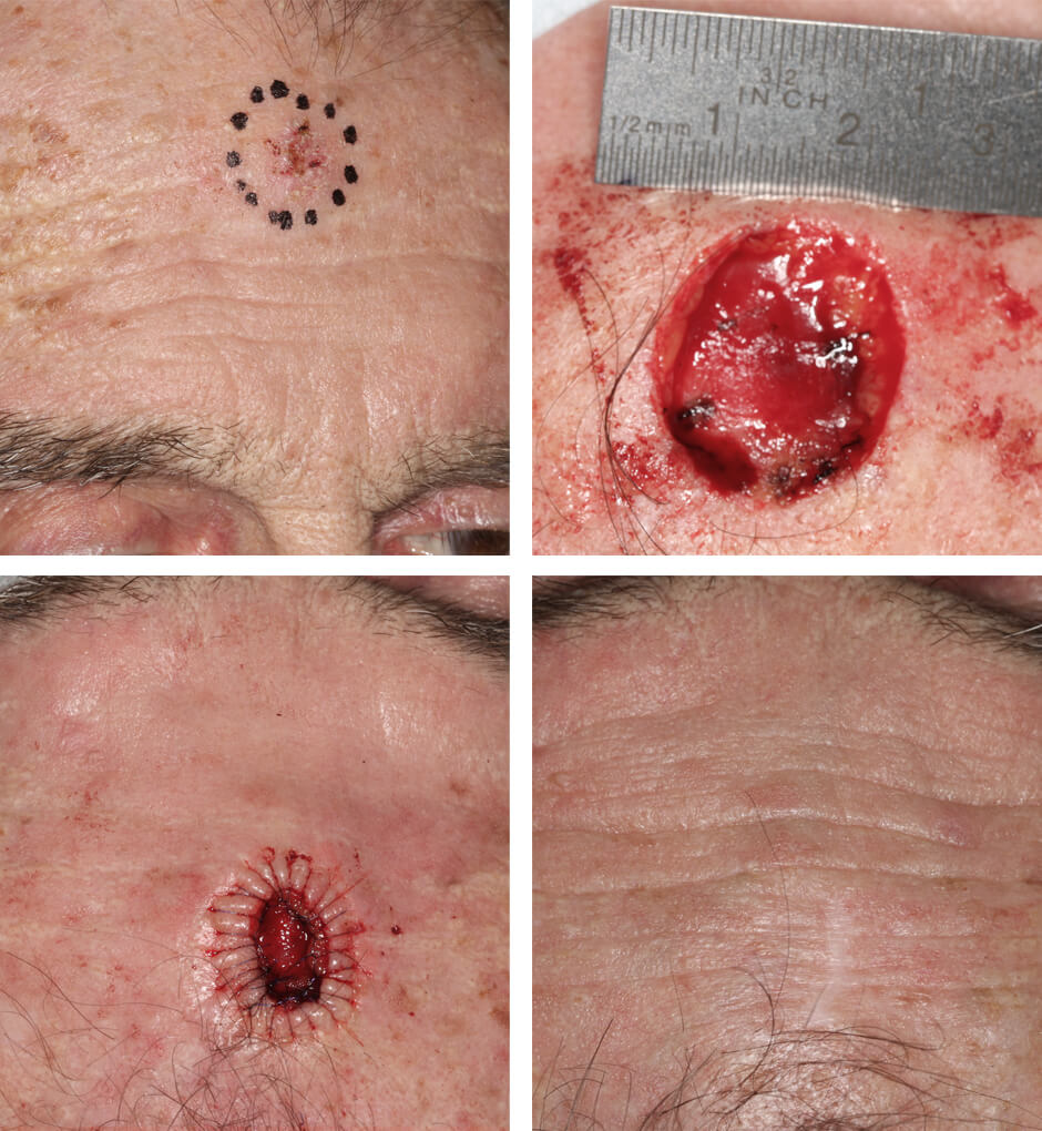
Fig. 1. Non-melanoma skin cancer on the forehead with the resection area marked.
Fig. 2. Skin defect with ruler.
Fig. 3. Circumferential purse-string suture.
Fig. 4. Final result.
Content warning: The following images contain photographs of a wound.
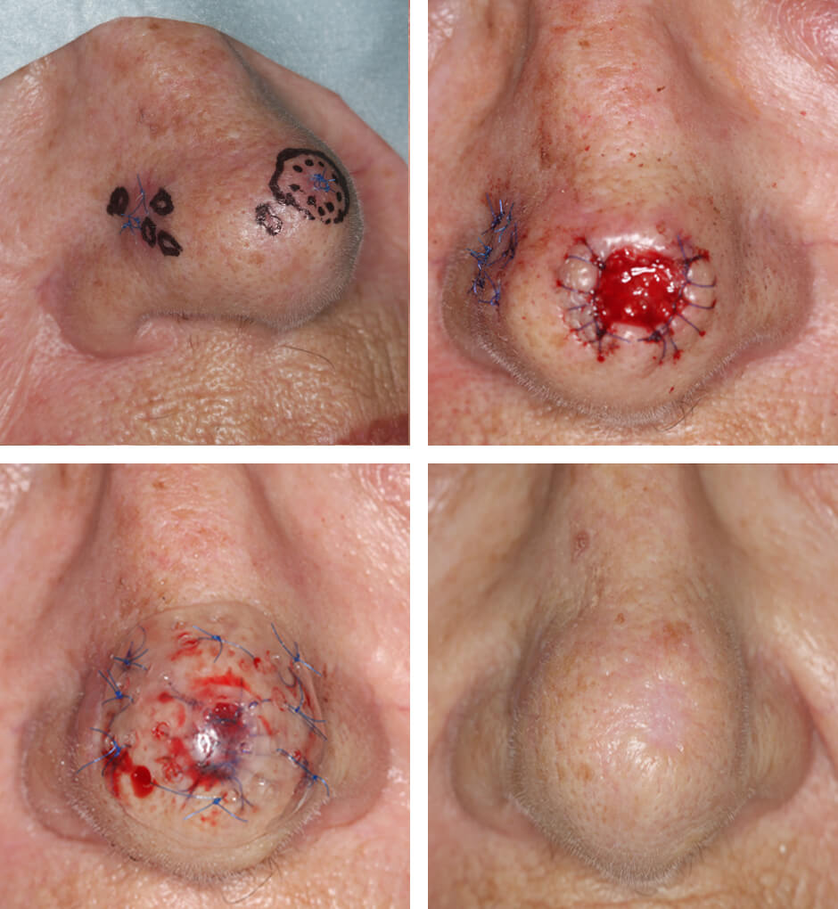
Fig. 1. Resection line marked on the nasal tip. Additional samples were taken from the right side but were found to be tumor-free.
Fig. 2. Circumferential purse-string suture.
Fig. 3. Wound plate sutured in place.
Fig. 4. Final result.
Content warning: The following images contain photographs of a wound.
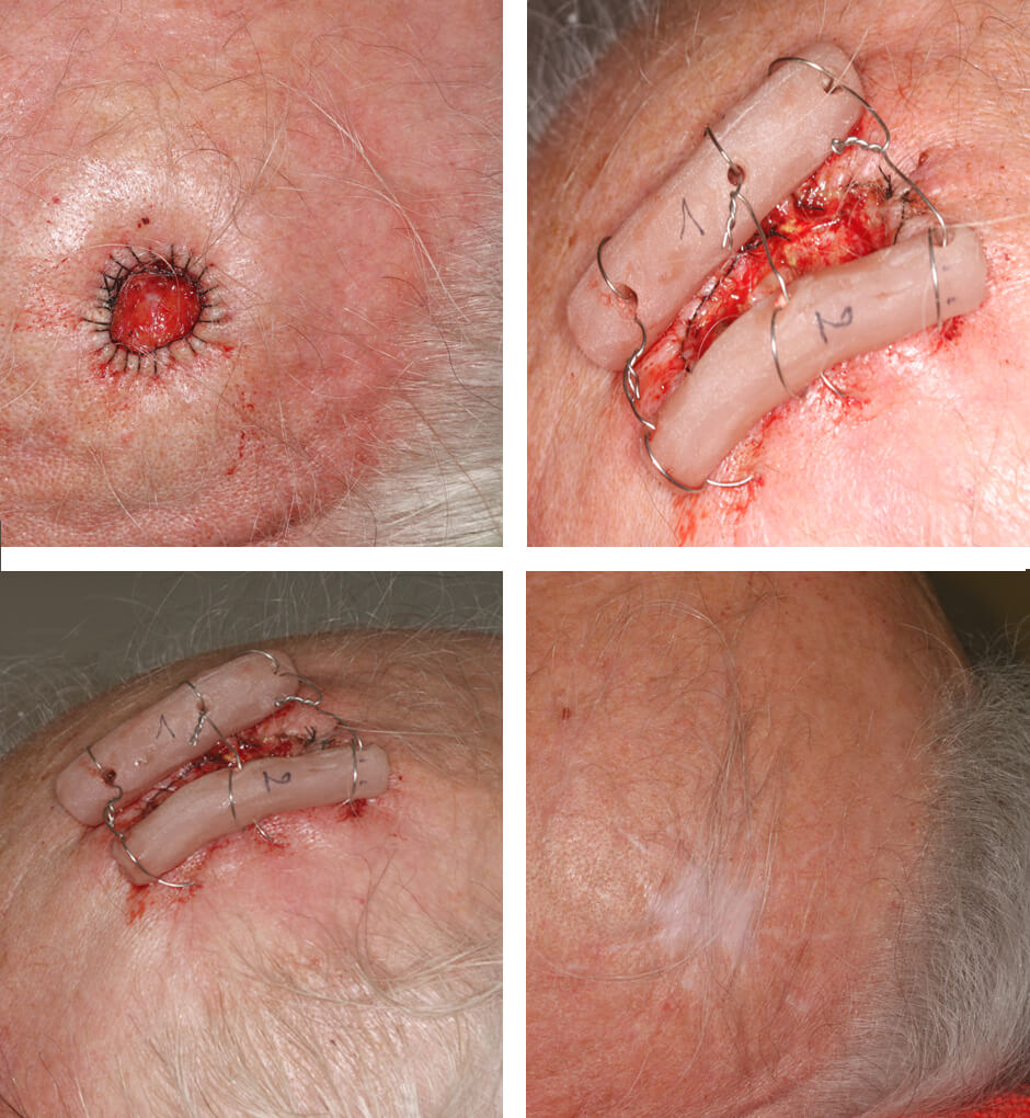
Fig. 1. Scalp defect with circumferential purse-string suture.
Figs. 2–3. Wire cerclages with plastic pulleys to prevent tearing of the skin.
Fig. 4. Final result.
Content warning: The following images contain photographs of a wound.
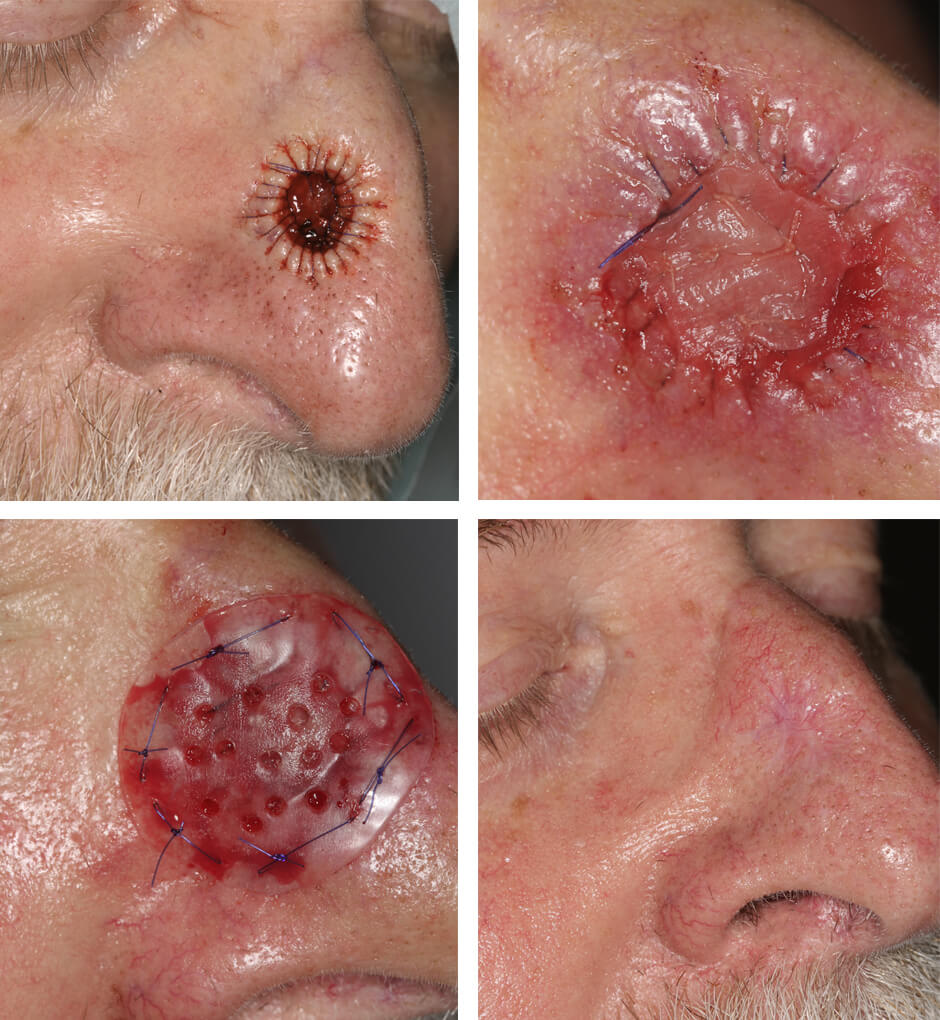
Fig. 1. Defect on the right side of the nose with circumferential purse-string suture.
Fig. 2. Wound covered with a resorbable membrane.
Fig. 3. Wound plate sutured in place.
Fig. 4. Final result.
(Preauricular Region)
Content warning: The following images contain photographs of a wound.
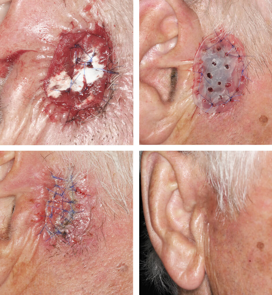
Fig. 1. Wound covered with a resorbable membrane. The anterior edge of the right ear is visible on the left side of the image.
Fig. 2. Wound plate sutured in place.
Fig. 3. Additional circumferential purse-string suture.
Fig. 4. Final result.
Content warning: The following images contain photographs of a wound.
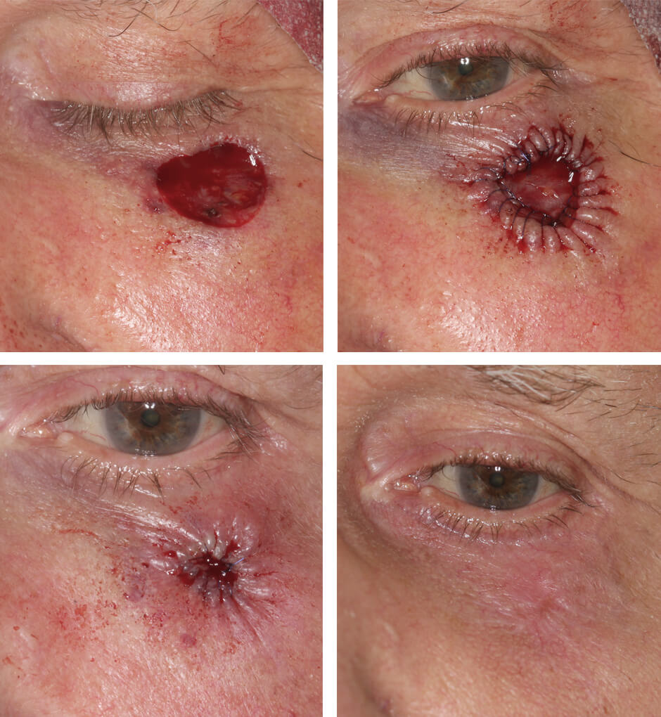
Fig. 1. Defect in the left eyelid.
Fig. 2. Circumferential purse-string suture.
Fig. 3. Additional circumferential purse-string suture.
Fig. 4. Final result.
How to Interpret Results Realistically
Results depend, among other factors, on:
- The size and location of the original wound
- Individual skin characteristics
- The healing process
- Personal factors
The goal of treatment is not cosmetic perfection, but rather a medically safe and functionally optimal outcome.
Medical Information: Dr. med. Dr. med. dent. Andreas Born