
A diagnosis of skin cancer on the face can naturally be unsettling at first. In addition to concerns about your health, another question often arises quickly: What will the treatment mean for my face, my appearance, and my daily life?
The good news is:
Many forms of skin cancer can now be treated very effectively if they are completely removed. At the same time, there are gentle surgical procedures that can help avoid large scars and extensive plastic surgery, especially in the sensitive facial and scalp areas. We’d like to provide you with more information about this on the following pages.
Are you affected by skin cancer on the face?
Below you will find some basic information on the topic:

Categorizing Skin Cancer:
What is meant by “skin cancer”
Not every skin change is dangerous, and not every form of skin cancer progresses in the same way. A medical evaluation is always essential. The term “skin cancer” encompasses various malignant skin diseases that differ in their behavior, extent, and treatment.
1. Basal Cell Carcinoma (BCC) (synonym: Basalioma)
is a form of non-melanoma skin cancer which accounts for approximately 80 % of all skin cancers in the head and neck region, generally does not spread to other parts of the body; however, it is “malignant” in its local behavior, as it infiltrates and destroys surrounding healthy tissue. The tendency to send tumor extensions into neighboring tissue is characteristic of white skin cancer and requires a more extensive excision than the skin lesion visible to the naked eye. Basal cell carcinomas very rarely form metastases; however, they can invade and destroy the surrounding healthy tissue at the site of origin.
2. Melanoma
We also treat melanoma in its early stage (lentigo maligna)—when there is not yet any indication of metastatic spread —in collaboration with dermatologists.
3. Squamous Cell Carcinoma
Squamous cell carcinomas of the skin on the face, head, or neck typically occur in older adults over the age of 70. This type of cancer also has the ability to metastasize, although metastases initially tend to occur in the lymph nodes along the lymphatic drainage pathways. However, this is extremely rare when the tumor is smaller than 2 cm.
For more extensive squamous cell carcinomas, further diagnostic testing of potentially affected lymph nodes or organs—known as staging—may therefore be necessary prior to surgery. We can arrange this in collaboration with radiology, laboratory medicine, and other specialist practices.
Why Complete Removal Is Important
Skin cancer develops when skin cells multiply uncontrollably and invade neighboring skin or even organs. The body can no longer repair this damage on its own. To achieve the best possible chance of a full recovery, it is therefore important to remove the tumor completely. This also helps reduce the risk of the skin cancer reappearing in the same place—a so-called recurrence.
Treatment Methods for Skin Cancer
Non-surgical Methods
Depending on the type and extent of the skin cancer, non-surgical treatments may also be considered, such as topical ointments and creams, laser and photodynamic therapy, radiation therapy, or other drug-based treatments involving pills or chemotherapy. Since these treatments are not offered at our practice, they will not be discussed in detail here.
Surgical Procedures
We prefer a surgical procedure in which the affected tissue is completely removed (excision). During and/or after the procedure, the removed tissue is examined under a microscope by pathologists (histology, Figure 1). The histological examination determines whether the cancer has been completely removed.
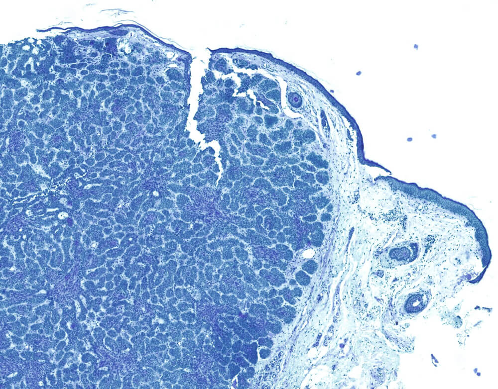
Figure 1.: What looks like an interesting pattern or a modern painting is actually a microscopic (histological) image of Basal Cell Carcinoma. The cancer cells are visible in the left and center portions of the image. They can be identified by the numerous blue-stained cell nuclei.
Source: Institute of Pathology at Marienhospital Stuttgart, Germany
Not only is the visibly altered tissue removed, but also a margin of “apparently healthy” skin surrounding the tumor. This so-called safety margin ensures that even the smallest traces of cancer—which are not visible to the naked eye—are removed. This is why it is also referred to as a necessary safety margin. Microscopic/histological examination by pathologists is essential for the success of treatment.
Particularly for the common forms of non-melanoma skin cancer, it is recommended to maintain a safety margin of approximately 3 mm around the tumor whenever possible. Ultimately, the decisive factor is the histological examination, which verifies whether the tumor has been completely removed.
This approach may differ for melanoma and squamous cell carcinoma. It is always adapted to the specific medical situation and thus contributes significantly to the often very good chances of cure for many forms of skin cancer.
In this context, the term “oncological radicality” is used. This means that tissue is removed until the tumor is completely eliminated. For the very common white skin cancer (also known as basal cell carcinoma), the chances of cure are as high as 95% when the surgery is performed correctly. The prognosis is similarly good for an early form of melanoma (lentigo maligna) as well as for squamous cell carcinoma when it is smaller than 2 cm.
Chances of Recovery – A Realistic Assessment
Many forms of skin cancer can be treated very effectively today, especially if they are detected early and completely removed. That is why regular skin cancer screening is so important for successful treatment.
If skin cancer is suspected, a thorough examination should be conducted by doctors who specialize in this field. If there is a specific suspicion of skin cancer, a tissue sample is usually taken and then examined under a microscope (see above).
An individual’s chances of recovery depend, among other things, on the type of skin cancer, its size and location, and the timing of the diagnosis.
Why Skin Cancer on the Face Poses Special Challenges
If the skin cancer is located on the face—for example, on the nose, eyelids, or scalp—the treatment poses additional challenges. These areas are highly visible and functionally sensitive.
Treatment must therefore combine two goals: the medically safe removal of the tumor and an approach that is as gentle as possible on healthy tissue while preserving function and appearance.
The type of wound closure and the subsequent healing process are correspondingly important.
The method of wound closure and the subsequent healing process are therefore particularly important.
Sources:
Frerich, B., Prall, F. (2018): Basalzellkarzinome der Gesichts- und Kopfhaut. Update der Therapieverfahren. Der MKG-Chirurg 11: 49-63
Lang, B. et al. (2024): S2k-Leitlinie Basalzellkarzinom der Haut (Aktualisierung 2023). https://opendata.uni-halle.de/bitstream/1981185920/120126/1/J Deutsche Derma Gesell – 2024 – Lang – S2k‐Leitlinie Basalzellkarzinom der Haut Aktualisierung 2023.pdf
What to Expect Next
On the next page, you’ll find information about a gentle alternative to traditional surgery: wound contraction for skin cancer on the face
Medical Information: Dr. med. Dr. med. dent. Andreas Born